Bone Pathology in Equine Wobbler Syndrome presented at 2021 UK Equine Research Showcase
 University of Kentucky hosted the first session of its 10th annual UK Equine Research Showcase in a virtual setting Jan. 5. The session emphasized musculoskeletal topics in weanling to yearling horses and presented both completed and work-in-progress projects.
Jennifer Janes, DVM, PhD, Dipl. ACVP, associate professor of anatomic pathology at UK’s Veterinary Diagnostic Laboratory presented on bone pathology in equine Wobbler Syndrome during the session.
She focused on the condition seen in younger horses, which can develop anywhere from 6 months to 7 years of age depending on breed.
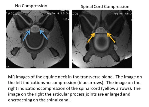
Janes defined equine Wobbler Syndrome as an equine neurological disease resulting from spinal cord compression in the neck due to vertebral malformations. This is a disease that is caused by skeletal malformations or related pathological changes that decrease the space available in the spinal canal. On a clinical level, it presents as a neurological disorder. The underlying skeletal changes that lead to a stricture or narrowing of the spinal canal can be variable. What they all have in common, however, is a resulting compression of the spinal cord that leads to the observed neurological deficits.
According to Janes, research shows that although the disease isn’t gender specific, males are more predisposed to developing wobblers compared to females, by a ratio anywhere from 3:1 up to 15:1 described in the literature. The disease is seen most commonly in Thoroughbreds, Tennessee Walking Horses and Warmbloods, but can be found in other breeds.
According to Janes, neurologic deficits are typically more severe in the hind limbs than the forelimbs in Wobblers. This is because nerve tracts that control hind limb placement are more superficial in the spinal cord. Thus, they are the first to be a compressed due to vertebral malformations
Janes said Wobbler Syndrome is considered a multifactorial disease with contributing factors including rapid growth, high energy diets and altered copper and zinc. A potential genetic role is suspected but has yet to been specifically characterized. Available treatment options range from conservative management and nutritional changes to surgical intervention. Appropriate treatment recommendations can be made in consultation with a horse owner’s veterinarian.
There is evidence showing that horses with Wobbler Syndrome can have increased frequency of osteochondrosis in the neck as compared to unaffected horses. Osteochondrosis is a developmental orthopaedic disorder where the normal transition of cartilage to bone does not occur. In Wobblers, osteochondrosis is located in the articular processes of the cervical vertebrae.
Janes and colleagues investigated articular process pathology in the entire cervical column, comparing horses with Wobblers Syndrome to unaffected horses. The goal was to increase knowledge on the skeletal pathology associated with the disease in order to advance our understanding of the underlying causes and disease mechanisms.
As background, according to Janes, articular process joints in the neck are synovial joints that function to link adjacent vertebrae in the column. For reference, the knee is a type of synovial joint. This type of joint is composed of two adjacent bones lined by articular cartilage that are connected by a joint capsule and synovial fluid fills the intervening joint space.
The investigative approach was to first quantitatively assess lesions identified on postmortem MRI (magnetic resonance imaging). Secondly, a representative group of identified of articular process bone and cartilage lesions were further characterized using micro-CT (computerized tomography) and histopathology.
Findings included cartilage and bone lesions in the articular processes occurred with more frequency in Wobbler horses as compared to controls. In addition, articular process lesions were not limited to only sites of compression but also located at sites away from compression as well. All lesions involving the articular process cartilage were osteochondrosis. Lesions in the bone included bone cysts, areas of fibrosis and osteosclerosis (thickening of the bone).
“Osteochondrosis and true bone cysts support developmental aberrations in bone and cartilage maturation and osteosclerosis was also observed, supporting likely secondary biomechanical influences,” Janes said.
Katelynn Krieger, a junior majoring in equine science and management, is a communications and student relations intern with UK Ag Equine Programs.
